


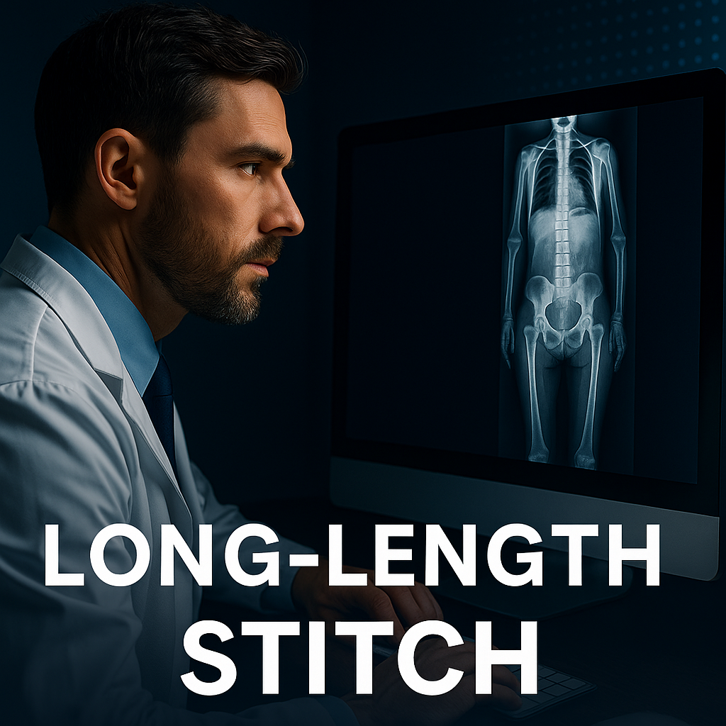
Long-Length Stitch in DICOM Viewers: Why It Matters and How XEUS Makes It Better
Introduction
In modern diagnostic imaging, accuracy often depends not only on the quality of each individual exposure but also on the ability to see the entire anatomy as one continuous structure. This is especially true in orthopedics, spinal assessments, scoliosis follow-ups, leg-length evaluations, and pre-operative planning. Because the human spine or lower extremities are longer than the detector size of most X-ray systems, they require multiple exposures—typically overlapping segments.
This is where the Long-Length Stitch feature in DICOM viewers becomes essential. Long-Length Stitching (also known as Full Spine / Full Leg Reconstruction) automatically merges multiple radiographic images into one seamless, high-resolution long image.
What Is Long-Length Stitching?
Long-Length Stitching is the process of combining multiple X-ray images—usually acquired in overlapping sections—into a single, unified long-format image. Typical examples include:
-
Full Spine (S-Curve evaluation, scoliosis measurement)
-
Full Leg (hip–knee–ankle alignment)
-
Lower-limb discrepancy studies
-
Orthopedic surgical planning
Because detectors are smaller than the entire area of interest, stitching ensures that clinicians can analyze the complete anatomy without missing transitional zones.
Why Long-Length Stitching Matters in Clinical Practice
1. Full Anatomical Context
Clinicians need to see the entire structure—not isolated segments. Stitching provides a complete, continuous view, which is critical for:
-
Cobb Angle measurement
-
Long-leg mechanical axis assessment
-
Tracking deformities
-
Pre- and post-surgical evaluation
2. Higher Diagnostic Confidence
With a unified stitched image, radiologists can precisely follow curves, joint positions, or alignment issues along the entire length of the spine or limbs.
3. Eliminating Gaps & Overlapping Errors
Modern stitching algorithms remove:
-
intensity differences,
-
magnification distortions,
-
geometric warping, and
-
overlapping misalignment.
This ensures a clean, consistent final image.
4. Better Surgical Planning
Orthopedic surgeons rely heavily on accurate stitched images for:
-
guiding osteotomy
-
planning limb reconstruction
-
selecting implants
-
monitoring recovery progress
How Long-Length Stitch Works (Technical Overview)
Although different DICOM viewers have distinct implementations, the workflow is generally similar:
Step 1: Importing the Stitch Series
Stitch studies are usually identified by:
-
tagging (e.g., “stitch,” “full spine,” “full leg”),
-
acquisition protocols, or
-
consecutive series with overlapping anatomy.
Step 2: Detecting Overlaps
Algorithms analyze overlapping regions between images using:
-
pixel intensity matching
-
gradient alignment
-
anatomical landmark recognition
Step 3: Exposure Normalization
Because exposure varies between segments, the system applies:
-
brightness/contrast equalization
-
histogram matching
-
blending masks
Step 4: Seamless Merging
Images are merged using:
-
linear blending
-
multi-band blending
-
edge-aware smoothing
to produce a seamless long image.
Step 5: Final Reconstruction
The output is a high-resolution long-format DICOM ready for:
-
measurement tools
-
reporting
-
exporting
-
printing
Clinical Applications of Long-Length Stitching
Scoliosis Evaluation
The stitched image allows measurement of:
-
Cobb angle
-
vertebral rotation
-
kyphosis and lordosis
-
progression over time
Pediatric Orthopedics
Essential for early detection of:
-
congenital deformities
-
limb-length discrepancies
-
growth abnormalities
Trauma & Sports Medicine
Monitoring long-bone fractures, alignment, and recovery.
Preoperative Planning
A must-have for full-leg alignment and designing surgical intervention.
Challenges in Stitching (and Why Good Software Matters)
Despite its usefulness, stitching is not trivial. Challenges include:
-
Patient movement during acquisition
-
Slight changes in positioning
-
Detector distortion
-
Exposure differences between panels
-
Soft tissue variation
A professional stitching engine must address all these issues with minimal manual correction.
Long-Length Stitch in XEUS: Smarter, Faster, Cleaner
XEUS by MiNNOVAA takes the Long-Length Stitch feature to a higher level by focusing on accuracy, speed, and clinical usability.
1. Intelligent Auto-Stitch Algorithm
XEUS automatically detects overlap and aligns images with sub-pixel precision.
This reduces the need for manual adjustments and guarantees consistent results.
2. Exposure & Intensity Equalization
The engine applies dynamic normalization to ensure uniform brightness across all segments.
No more harsh lines or exposure jumps.
3. Full-Length Image Optimization
High-fidelity reconstruction preserves:
-
bone edges
-
joint landmarks
-
spinal morphology
This is crucial for orthopedic accuracy.
4. Seamless Integration with Tools
Stitched images in XEUS are fully compatible with:
-
Cobb Angle
-
Leg-length measurements
-
Multi-point line tools
-
Angle & Axis evaluation
-
TID-1500 structured reporting
5. Fast & User-Friendly Workflow
The user simply selects the series and clicks “Stitch” —
XEUS does the rest automatically.
6. Designed for Real Clinical Use
Whether it’s pediatric scoliosis follow-up or adult full-leg planning, XEUS ensures radiologists and surgeons always have a clean, accurate stitched image to rely on.
Conclusion
Long-Length Stitching is not just a convenience feature—it is a clinical necessity. From orthopedic surgeries to scoliosis evaluation, having a unified full-length radiographic image dramatically improves diagnostic precision and patient care.
With XEUS, stitching becomes:
faster, sharper, smarter, and beautifully seamless.
A single click delivers a full-length, clinically accurate image ready for diagnosis, reporting, and printing.