In the world of medical imaging, DICOM (Digital Imaging and Communications in Medicine) is the gold standard for managing and sharing image data. What sets DICOM apart from other image formats is its ability to store not just images but also a wealth of associated information, including detailed patient data, study descriptions, and image acquisition parameters. This blog post will delve into the rich data contained in DICOM files, explaining what information is stored, the attributes that can be extracted, and how this data is used in medical imaging.
What is a DICOM File?
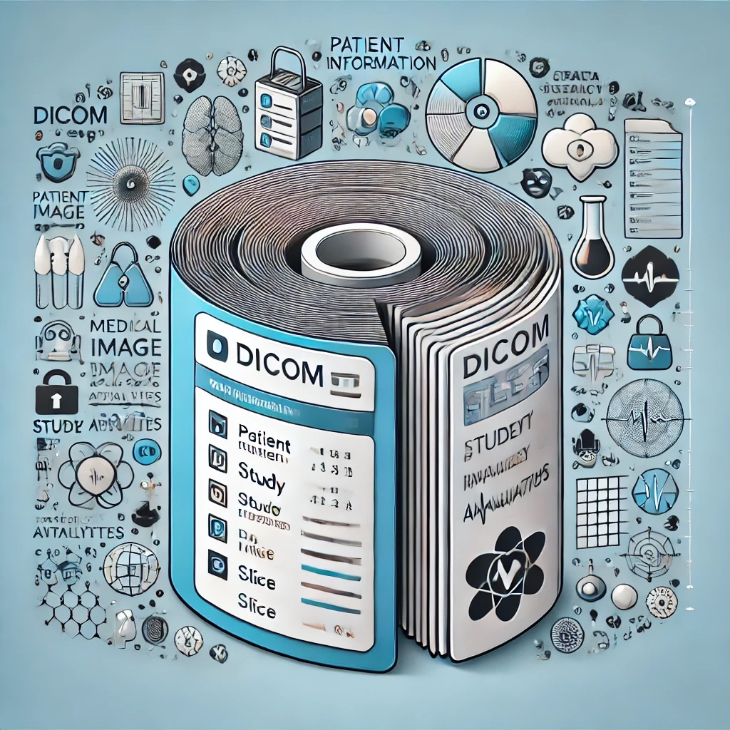
A DICOM file is more than just an image; it is a complex container that holds both the visual data and a comprehensive set of metadata. This metadata provides context to the image, making it possible to interpret, analyze, and use the image effectively within the healthcare system. The combination of image data and metadata in a single file ensures that the image can be accurately associated with the correct patient, study, and diagnostic process.
Key Components of a DICOM File
-
Image Data
The primary component of a DICOM file is the image data itself. This could be a single image or a series of images, such as slices from a CT scan or frames from an ultrasound study. The image data is stored in a standardized format, allowing it to be displayed consistently across different DICOM viewers and systems.
-
Patient Information
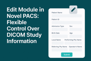
DICOM files contain detailed patient information, which is crucial for ensuring that the images are correctly linked to the right patient. This information typically includes:
- Patient Name: The full name of the patient.
- Patient ID: A unique identifier for the patient within the healthcare system.
- Date of Birth: The patient’s date of birth, which can be important for age-related diagnoses.
- Sex: The patient’s gender, which can be relevant for certain types of medical imaging and diagnosis.
- Patient Address and Contact Information: While not always included, this information can be part of the DICOM file, especially in systems where detailed patient records are maintained.
-
Study Information
In addition to patient information, DICOM files also contain data about the imaging study itself. This includes:
- Study Date and Time: The date and time when the imaging study was performed.
- Study Description: A textual description of the study, often provided by the radiologist or technician.
- Study Instance UID: A unique identifier for the study, ensuring that it can be accurately referenced within the healthcare system.
- Referring Physician: The name of the physician who ordered the imaging study.
- Accession Number: A unique number assigned to the imaging study, used to track it within the hospital or clinic.
-
Series Information
DICOM files also organize images into series, with each series representing a specific acquisition or phase of the study. The series information includes:
- Series Number: A unique number assigned to the series within the study.
- Modality: The type of imaging modality used (e.g., MRI, CT, X-ray).
- Series Description: A textual description of the series, often indicating the specific area of the body or the technique used.
- Series Instance UID: A unique identifier for the series.
-
Image Attributes
One of the most critical aspects of DICOM is the extensive set of attributes that describe the images themselves. These attributes provide detailed information about how the images were acquired, processed, and stored. Some key image attributes include:
- SOP Class UID: Identifies the type of DICOM service object pair (SOP) class, such as a specific imaging modality or treatment plan.
- Pixel Data: The raw data for the image pixels, including the number of rows and columns, bit depth, and pixel spacing.
- Photometric Interpretation: Indicates how the pixel data should be interpreted, such as grayscale or RGB color.
- Window Center and Width: These values define the brightness and contrast settings for the image, which are crucial for interpreting medical images.
- Slice Thickness and Spacing: For modalities like CT and MRI, this attribute defines the thickness of the slices and the spacing between them.
- Acquisition Parameters: Information about the imaging device settings, such as the exposure time, kVp (kilovolt peak), and mAs (milliampere-seconds), which are important for understanding the quality and characteristics of the image.
Extracting and Using DICOM Data
The wealth of information contained in DICOM files can be leveraged in various ways to enhance medical imaging workflows and improve patient care. Here’s how this data is used:
-
Image Display and Interpretation
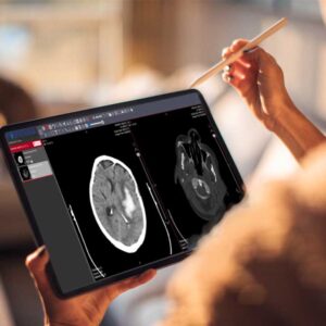
The attributes stored in a DICOM file are essential for accurately displaying the image. For example, the window center and width settings ensure that the image is displayed with the correct brightness and contrast, allowing radiologists to see the details they need to make a diagnosis. Similarly, the pixel spacing and slice thickness attributes help in reconstructing the images in 3D or in generating accurate measurements.
-
Patient and Study Management
The patient and study information stored in DICOM files allows for seamless integration with other healthcare IT systems, such as electronic health records (EHRs) and radiology information systems (RIS). This integration ensures that images are correctly associated with the patient’s medical record, reducing the risk of errors and improving the efficiency of the imaging workflow.
-
Research and Analysis
The detailed metadata in DICOM files makes them valuable for research and analysis. Researchers can use DICOM data to study imaging techniques, analyze disease patterns, and develop new diagnostic tools. The ability to extract specific attributes from DICOM files also facilitates large-scale studies that require the analysis of thousands of images.
-
Quality Assurance
DICOM files play a crucial role in quality assurance (QA) within medical imaging departments. By analyzing the acquisition parameters and image attributes, QA teams can ensure that imaging devices are functioning correctly and producing images of the required quality. This data can also be used to track trends and identify potential issues before they affect patient care.
-
Advanced Image Processing
Advanced image processing techniques, such as 3D reconstruction, image fusion, and artificial intelligence (AI) algorithms, rely on the detailed attributes stored in DICOM files. For example, 3D reconstruction software uses slice thickness and spacing data to create accurate 3D models, while AI algorithms may use pixel data and photometric interpretation to identify patterns and anomalies.
Conclusion
DICOM files are much more than simple image containers; they are comprehensive data packages that include a wide range of information about the patient, study, and image attributes. This rich metadata enables healthcare professionals to accurately interpret images, manage patient data, conduct research, and ensure the quality of imaging services.
As the field of medical imaging continues to evolve, the ability to extract and utilize the data contained in DICOM files will become increasingly important. Whether it’s through advanced image processing techniques, AI integration, or web-based applications like DICOMweb, the future of medical imaging will be shaped by how effectively we can leverage the data stored in these powerful files.
In future blog posts, we will explore how DICOM files contain detailed patient information and the implications this has for patient care, data security, and interoperability. Stay tuned for more insights into the world of medical imaging and healthcare technology.


