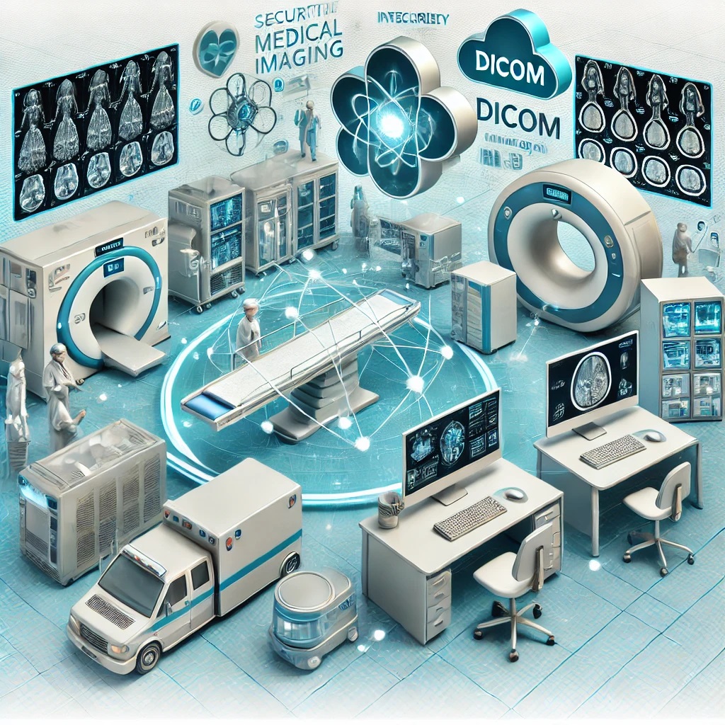
In the rapidly evolving field of medical technology, the efficient and accurate exchange of information is crucial. Digital Imaging and Communications in Medicine, commonly known as DICOM, is a cornerstone of this process. DICOM is not just a standard; it is the lifeline that allows different medical imaging devices and systems to communicate seamlessly. This blog post will delve into what DICOM is, its history, key parameters, features, recent innovations, and complementary protocols that enhance its functionality.
What is DICOM?
DICOM is a global standard for handling, storing, printing, and transmitting information in medical imaging. It includes a file format definition and a network communications protocol. DICOM files can contain a wide range of data, including patient information, medical images, and metadata describing how the images were obtained and processed.
The importance of DICOM cannot be overstated, as it ensures interoperability between different imaging devices such as MRI, CT, and X-ray machines, regardless of the manufacturer. This interoperability is vital in medical environments where images from multiple sources must be integrated and analyzed together.
History of DICOM
The journey of DICOM began in the early 1980s when the American College of Radiology (ACR) and the National Electrical Manufacturers Association (NEMA) collaborated to develop a standard for the electronic transmission of medical images. The first version, ACR/NEMA 300, was released in 1985 but had limited success due to its lack of network capabilities.
DICOM 3.0, released in 1993, was a game-changer. It introduced network support, which allowed different systems to communicate over local and wide-area networks. This version also standardized the image format, ensuring that images from different devices could be viewed and analyzed on any DICOM-compatible system. Since then, DICOM has undergone several revisions, each adding new features and capabilities to keep pace with advancements in medical imaging technology.
Key Parameters in DICOM
Several key parameters in DICOM are critical for ensuring the standard’s functionality and interoperability:
-
Modality: This parameter identifies the type of equipment that generated the images, such as CT, MRI, or ultrasound. It ensures that the images are correctly interpreted according to their source.
-
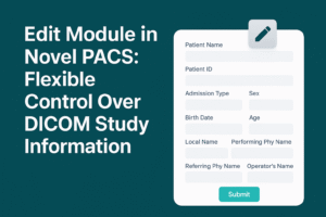
Patient Information: DICOM files contain detailed patient information, including name, age, sex, and medical history. This metadata is essential for associating images with the correct patient and ensuring continuity of care.
-
Image Attributes: DICOM defines a wide range of attributes for images, including resolution, bit depth, and pixel spacing. These attributes ensure that images are displayed correctly and consistently across different systems.
-
Image Compression: DICOM supports various compression algorithms, including lossless and lossy compression, to reduce the size of image files without compromising diagnostic quality.
-
Networking Protocols: DICOM uses the TCP/IP protocol for network communication, enabling the transfer of images and associated data between different systems.
Features
DICOM offers several features that have made it the de facto standard in medical imaging:
-
Interoperability: One of DICOM’s primary features is its ability to ensure that different imaging devices and systems can communicate and share data seamlessly. This interoperability is vital for modern healthcare, where imaging data often needs to be shared across departments and institutions.
-
Extensibility: it is designed to be extensible, meaning that new features and capabilities can be added without disrupting existing systems. This flexibility has allowed DICOM to evolve alongside advancements in medical imaging technology.
-
Data Security: it includes features for ensuring the security and confidentiality of patient data. This includes support for encryption and digital signatures, which help protect data during transmission and storage.
-
Integration with PACS: DICOM is closely integrated with Picture Archiving and Communication Systems (PACS), which are used to store, retrieve, and manage medical images. This integration allows for efficient workflows and easy access to imaging data across the healthcare enterprise.
-
Compatibility with Other Standards: it is designed to work alongside other medical standards, such as HL7, which is used for exchanging clinical and administrative data. This compatibility ensures that DICOM can be integrated into a broader healthcare IT environment.
Recent Innovations
DICOM has continued to evolve in response to advancements in medical imaging technology and changing healthcare needs. Some of the latest innovations in DICOM include:
-
DICOMweb: DICOMweb is a set of web-based protocols that allows DICOM data to be accessed and exchanged over the internet using standard web technologies like HTTP and RESTful APIs. This makes it easier to integrate DICOM with web-based applications and cloud-based services, facilitating remote access to medical images.
-
AI Integration: With the rise of artificial intelligence in healthcare, there has been a growing interest in integrating AI algorithms with DICOM. This integration allows AI tools to directly access DICOM data for tasks like image analysis, enhancing the diagnostic capabilities of medical imaging systems.
-
Improved Compression Techniques: Recent updates to DICOM have introduced more advanced compression techniques, such as JPEG 2000, which offer better image quality at reduced file sizes. This is particularly important in the era of high-resolution imaging, where file sizes can become very large.
-
Enhanced Metadata Support: Newer versions of DICOM have expanded the range of metadata that can be included with images. This includes support for additional clinical information, such as genetic data and patient outcomes, which can be used to provide a more comprehensive view of the patient’s health.
Complementary Protocols to DICOM
While DICOM is a comprehensive standard, it is often used in conjunction with other protocols to provide a complete solution for medical imaging and healthcare IT. Some of these complementary protocols include:
-
HL7 (Health Level Seven): HL7 is a set of standards for the exchange of clinical and administrative data between healthcare systems. It is often used alongside DICOM to ensure that imaging data can be integrated with other types of medical information, such as electronic health records (EHRs).
-
IHE (Integrating the Healthcare Enterprise): IHE is an initiative that promotes the use of established standards like DICOM and HL7 to improve the interoperability of healthcare systems. IHE profiles define how these standards should be used together to support specific clinical workflows, such as radiology or cardiology.
-
FHIR (Fast Healthcare Interoperability Resources): FHIR is a relatively new standard developed by HL7 for exchanging healthcare information electronically. It is designed to be more flexible and easier to implement than traditional HL7 standards, making it a good complement to DICOM in modern healthcare environments.
Conclusion
DICOM has been the backbone of medical imaging for decades, providing the standard that ensures different imaging devices and systems can communicate seamlessly. Its ability to evolve with technological advancements, support for a wide range of imaging modalities, and integration with other healthcare IT standards have made it indispensable in modern healthcare.
As DICOM continues to evolve, with innovations like DICOMweb and AI integration, it will remain at the forefront of medical imaging technology. Understanding DICOM is essential for anyone involved in the healthcare industry, as it is the key to unlocking the full potential of medical imaging.
In future posts, we will explore specific topics such as the role of JPEG 2000 in DICOM and how DICOM compares to other protocols commonly used in medical IT such as HL7. Stay tuned for more in-depth analysis and insights into the world of medical imaging and healthcare technology.
