Introduction
In modern medical imaging, three-dimensional visualization is essential for accurate diagnosis, treatment planning, and research. XEUS, a dedicated DICOM viewer for medical images, integrates advanced 3D tooling that turns volumetric datasets into interactive insight. With responsive navigation, curated 3D Presets, and precise Bounding Box controls, clinicians can focus on regions that matter and move from raw pixels to clinically useful views rapidly. This article reviews XEUS’s 3D feature set, how presets streamline routine work, and why bounding boxes are crucial for focused analysis and longitudinal follow-up.
1) The Role of the 3D Feature in XEUS
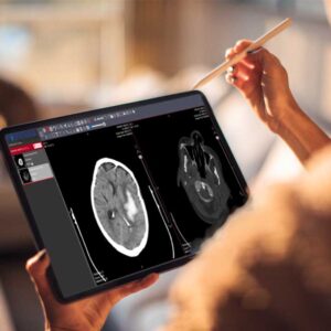
CT and MR are acquired as stacks of 2D slices. While conventional axial/coronal/sagittal views remain invaluable, they can obscure spatial relationships and small findings when viewed in isolation. XEUS’s 3D capability addresses this gap by reconstructing the volume into an interactive model:
- True volumetric context: View anatomy from arbitrary angles to understand adjacency (e.g., a lesion’s proximity to vessels, airways, or bony landmarks).
- Interactive control: Rotate, pan, zoom, and slice the model; peel away layers by adjusting opacity to reveal structures without losing orientation.
- Consistent geometry: Output respects the DICOM patient coordinate system, helping multi-disciplinary teams interpret the same structure coherently across sessions.
Technically, XEUS performs high-quality resampling from the source stack to render the volume as a coherent 3D object. This reduces stair-step artifacts, preserves edges, and keeps measurements made on 3D cross-sections consistent with in-plane tools. In practical use, a thoracic CT that shows faint nodularity on axial slices can, in 3D, reveal the nodule’s relationship to adjacent bronchovascular branches; a neuro case benefits from appreciating a mass effect relative to midline and ventricular systems.
2) 3D Presets in XEUS
3D Presets are predefined visualization recipes that quickly tune the 3D scene for a clinical task. Instead of adjusting multiple sliders manually, a preset sets typical parameters—such as transfer behavior, opacity ramps, light position, shading strength, cut-planes, and background—to reveal the target structures immediately.
Common categories include (examples vary by workflow):
- Bone-oriented: Accentuates high-attenuation structures for fracture, deformity, or pre-op templating.
- Soft-tissue emphasis: Balances parenchyma and vasculature for tumor assessment.
- Chest/vascular quick-look: Supports inspection of vessel paths and branching patterns.
- Brain parenchyma: Aids in appreciating mass effect or postoperative change with gentle shading.
Why presets matter:
- Speed: Jump from raw volume to a usable view in seconds.
- Consistency: Standardize how cases are reviewed within a team, improving inter-reader agreement.
- Customization: Start from a preset, then fine-tune contrast, brightness, clipping planes, and transparency for the patient at hand, and save your version for future use.
By reducing setup time, presets help clinicians stay focused on interpretation rather than wrestling with controls—particularly valuable in high-volume settings.
3) The Significance of the Bounding Box
The Bounding Box is a lightweight but powerful way to isolate a region of interest (ROI) within the 3D dataset. In XEUS, placing and adjusting a bounding box lets you:
- Focus analysis: Enclose a tumor, an arterial segment, or a bony corridor; suppress surrounding tissue to concentrate on the target.
- Improve visual clarity: Cropping reduces clutter and occlusion, so important edges and interfaces stand out.
- Track change over time: Reuse a comparable box across follow-up studies to visually and metrically compare the same anatomic region.
- Support segmentation steps: A cleanly bounded ROI is a practical precursor to manual or semi-automatic segmentation, improving speed and accuracy.
Controls are intuitive: drag faces, edges, or corners to resize; rotate to align the box with oblique anatomy; and lock the box when you’re ready to proceed. Because the bounding box constrains reslicing and projection, you can generate focused MPR/MIP views that align with the clinical question (e.g., stenosis grading or tumor margin inspection).
4) Enhancing Surgical Planning and Precision with 3D Imaging in XEUS
A key benefit of 3D in XEUS is pre-procedural planning. Whether for neurosurgery, orthopedics, ENT, or thoracic interventions, 3D views clarify approach, depth, and adjacency:
- Anatomic road-mapping: Rotate and clip to visualize safe corridors and anticipate obstacles (e.g., proximity to a sinus, foramen, artery, or nerve root).
- Target sizing and orientation: Use oblique cross-sections aligned with the long axis of a structure to derive true diameters/angles, minimizing bias from obliquity.
- Communication and documentation: 3D snapshots illustrate the plan clearly to colleagues and patients; presets ensure reproducible appearance across discussions.
By combining a relevant 3D Preset with a precisely placed Bounding Box, surgeons and interventionalists can focus the scene, simplify decision-making, and reduce surprises in the suite.
5) Challenges and Future Directions
While 3D visualization is powerful, there are practical considerations:
- Compute load: High-resolution volumes and advanced shading can be resource-intensive. XEUS mitigates this with efficient resampling and sensible defaults, but very large datasets may still demand robust workstations.
- Display discipline: Accurate perception depends on appropriate window/level and opacity settings; presets help, but complex cases may still need expert adjustment.
- Workflow integration: The best 3D view is only useful if it fits into the broader clinical workflow. Exporting views, sharing states, and keeping presentation consistent across readers are ongoing priorities.
- Looking ahead, broader use of GPU optimizations, smarter preset recommendations, and deeper interoperability with reporting systems will further streamline how 3D is used day-to-day.
Conclusion
XEUS brings together a capable 3D renderer, practical 3D Presets, and precise Bounding Box tools to turn volumetric data into actionable views. Clinicians can quickly orient themselves, isolate what matters, and communicate findings with clarity. By transforming 2D DICOM stacks into interactive 3D models—and by standardizing views through presets and ROI bounding—XEUS helps teams reach accurate diagnoses and confident plans faster. As rendering and
integration continue to advance, XEUS’s 3D feature set will only become more responsive and more central to high-quality clinical imaging.
for more information: https://academy.minnovaa.com/mod/page/view.php?id=18
References
3D / Volume Rendering
-
Radiopaedia — Volume rendering (overview and clinical context). Radiopaedia
-
Perandini S, et al. The diagnostic contribution of CT volumetric rendering techniques. Insights Imaging (open access). PubMed Central
-
Eid M, et al. Cinematic Rendering in CT: A Novel, Lifelike 3D Visualization Technique. AJR (review). ajronline.org
MPR / MIP (for cross-sectional & projection context)
-
Radiopaedia — Multiplanar reformation (MPR). Radiopaedia
-
Radiopaedia — Maximum intensity projection (MIP). Radiopaedia
-
Prokop M, et al. Use of maximum intensity projections in CT angiography. Radiographics (classic review). PubMed
-
Jerjir N, et al. MIP and MinIP in Practice (lungs; J Belg Soc Radiol, 2024). Belgian Radiology Journal
-
Xiang Z, et al. Diagnostic value of using MPR images (case-focused evidence). Pol J Radiol (PMC). PubMed Central
3D Presets / Transfer Presets
-
3D Slicer Docs — Volume rendering module (selecting/changing presets; user guide). slicer.readthedocs.io
-
3D Slicer Dev Docs — Volume rendering presets (preset files & configuration). slicer.readthedocs.io
Bounding Box / ROI Crop (workflow & tooling)
-
3D Slicer Docs — Crop Volume (cropping with an ROI “box” to focus a region). slicer.readthedocs.io
-
3D Slicer Script Repository — Get oriented bounding box & use Markups ROI. slicer.readthedocs.io
-
Slicer Forum (recent) — Creating a boundary box after rotating/translating a volume (practical notes). 3D Slicer Community
